Unique Microscope Installed in the Technology Building

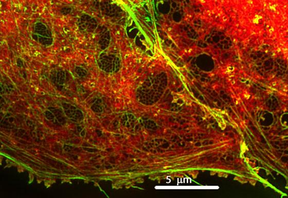 |
| The image above shows a surface cell from the liver with a unique level of detail. Nano-scaled pores are a part of the cell transport system and can be seen as black dots. The new SIM-microscope (structured illumination microscope) opens up a new world for researchers at UiT. |
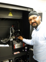 |
| The SIM microscope occupies as much space as 5 large refrigerators. In the picture above Balpreet demonstrates how cell samples are mounted onto the microscope. |
In Tromsø unique cell images can now be obtained thanks to the newly purchased microscope in the group of Balpreet Sing Ahluwalia at the Department of Physics and Technology. What differentiates this microscope from already existing models at UiT, is its ability to capture images of structures in the nano-scale region in for example liver cells. This instrument has an optical resolution of 20-50 nm, down to 0.000002 cm. The microscope is housed in the SIM-lab in the new Technology Building in Tromsø. The instrument was purchased for 6,5 million NOK from the US.
"As far as we know, this is the only instrument north of Oslo that can take images with nano-scale precision" says a proud Balpreet.
Close Cooperation Between Physicists and Molecular Biologists
Research on optics, sensor-technology and new applications of optical sensors in microscopy has brought physicists and molecular biologists together. Balpreet is working closely with specialists at the medical faculty, researching transport of compounds in the liver. The liver is in many ways the body's landfill removing harmful waste from the bloodstream. It also sorts out which medicines can pass through the body, and is therefore of great interest in medical research. With the new microscope, researchers can map out details in the liver transport system to a level that has never been seen before. For example, small wholes and pores central to transport of compounds in the liver can now be visualized.
Developing "Nano Filming" Technique
| Deanna Wolfson (left) and Cristina Øie interprets one of the very first images produced with the new instrument |
Two years ago, Balpreet Singh Ahluwalin received the prestigious ERC Starting Grant to develop a microscope able to film living cells. This film will allow researchers to study how the small pores in the liver cell are able to open and Close during cell transport. Studying the effect of medicine and alcohol at liver cells this new tool is expected to be essential. The technique is called "chip-based optical nanoscopy." A special chip for cell samples are developed and the optical properties of the chip enable visualization of detailed images of cell structures.
According to Balpreet, advanced microscopy today is done with very complex microscopes, while the cell samples are placed on a simple glass plate. Through his research Balpreet would like to do the opposite, using a simple microscope and an advanced chip for the sample.
"So far we have managed to confirm that the chip technology works, but still we need to obtain live images of transport in and out of the pores. This is a promising technology and we can not say much more because there are opportunities for patents here" , Balpreet says with a smile.
| PhD student Oystein Helle participates in the exciting work of developing chip-based optical nonoscopy. |
The facilities in the Technology building are part of the "Advanced Microscopy Core Facility" at the Faculty of Medicine and a resource that can be used by researchers throughout UiT.
Read More:
Prestisjefylt EU-støtte til UiT-forsker
Advanced microscopy core facility
|
Fact: Optical microscopes have a physical lower resolution of about 200-300nm. This is a fundamental lower limit surpassed only in recent years by new techniques. This resulted in a Nobel Prize to the field of optical nanoscopy (S. Hell 2014). The newly acquired OMX-microscope uses the SIM-technique (structure illumination microscopy) affording a resolution of about 100nm, and/or Single Molecule Localization Microscopy giving a resolution of ca 20-50nm. The latter technique gives good resolution but takes a lot of time |

