About the project
THERACAN is a research project focused on developing advanced theranostic strategies for personalized cancer treatment. The project combines diagnostic imaging and targeted radiotherapy in a single platform, aiming to improve both treatment precision and outcome monitoring. The project seeks to explore novel strategies to improve the efficacy of radioligand therapy including approaches for enhancing tumor targeting and retention of radiopharmaceuticals, personalizing protocols based on individual patient characteristics, tumor biology, and molecular profiles, and improved dosimetry procedures.
Work packages
WP-1 (Preclinical): Development of novel theranostic radioligands targeting elements of the TME displaying enhanced tumor specificity and tumor uptake
Main goal: Develop novel theranostic radioligands designed to target elements within the tumor microenvironment (TME)—such as PSMA and FAP—to bypass tumor cell heterogeneity, boost uptake, and extend tumor retention.
Key Activities: The team will optimize radiolabeling using therapeutic radionuclides (e.g., 64-Cu/67-Cu, 212-Pb, 225-Ac). In vitro assays, small animal PET/MRI dynamic imaging, and pharmacokinetic modeling will evaluate uptake and organ dosimetry. Vascular normalization strategies (focused ultrasound, low-dose EBRT) will also be explored to enhance radioligand delivery.

Main goal: Identify biomarkers predicting treatment response/resistance to targeted radionuclide therapy (TRT) and explore synergies with complementary treatments.
Key Activities: Utilizing tissue and serial blood samples from TRT-treated patients, researchers will analyze target expression, hypoxia markers, circulating tumor (ct)DNA, and extracellular vesicles (EVs). Immunophenotyping (via multiplex IHC and flow cytometry) will assess TRT-induced immune responses. Additionally, immunocompetent animal models will evaluate combined TRT strategies with immune checkpoint inhibitors and external beam radiotherapy (EBRT) to maximize systemic and anti-tumor efficacy.
WP-3 (Clinical): Patient selection for TRT treatments & clinical translation
Main goal: Translate preclinical innovations to clinical practice by personalizing TRT protocols using advanced whole-body imaging and novel dosimetry methods.
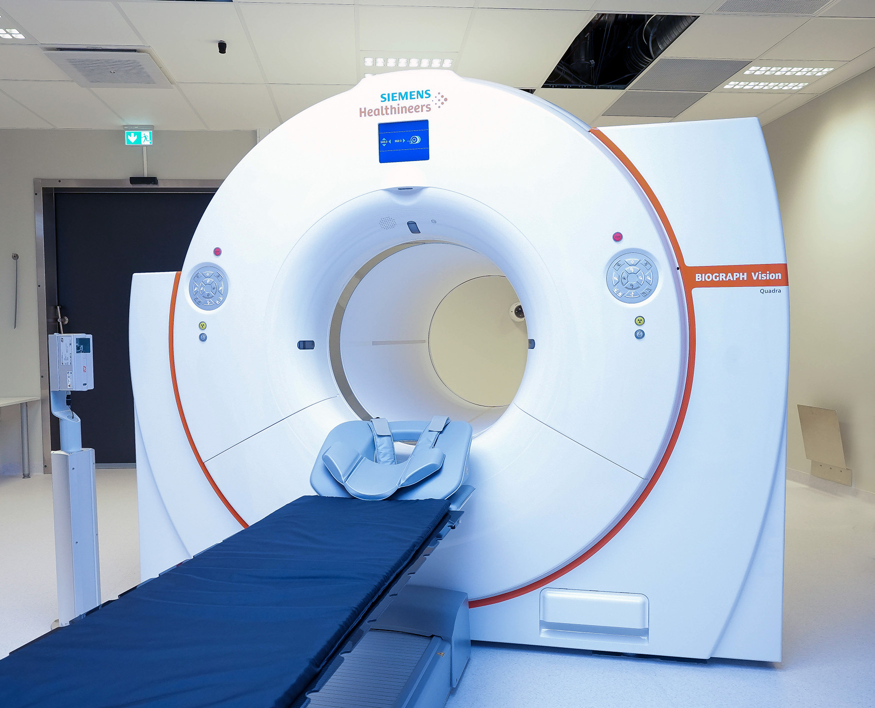
Key Activities: Leveraging a new Large-Axial-Field-of-View (LAFOV) PET/CT scanner, the team will perform dynamic whole-body tracer kinetic modeling to assess tumor retention. The package includes conducting compassionate treatments in selected advanced-stage cancer patients, evaluating standard-of-care cohorts, establishing non-invasive image-derived input functions, shortening scan protocols, and developing deep learning models for pretreatment internal dosimetry calculations.
Scientific approach & methodology
Radiotracer refinements: Development of new generation radioligands with increased affinity, increased in vivo stability and with higher killing potential.
Using molecular design and radiochemistry approaches, we will optimize the structure of targeting molecules —such as peptides and small molecules directed toward tumor-associated targets like PSMA and FAP —to improve how they behave in the body. By modifying key molecular features, we aim to increase tumor uptake, promote receptor-mediated internalization into cancers, and prolong retention of the radioactive compound within tumors. These improvements are expected to enhance the therapeutic effectiveness and safety of targeted radionuclide therapy and support development of more precise cancer treatments.
Novel approaches to enhance tumor uptake of radioligands, including ACT and LDRT
1: Focused ultrasound and Acoustic Cluster Therapy (ACT).
ACT involves the activation of microbubbles within tumor blood vessels using focused ultrasound, leading to a temporary and localized increase in vascular permeability. This effect enhances the delivery of targeted radiotherapeutics to the tumor tissue.
By improving tumor uptake, focused ultrasound increases both the therapeutic efficacy of the radiolabeled compounds and the accuracy of imaging-based dosimetry. This combination supports a more precise and individualized treatment approach.
2: Low dose radiotherapy (LDRT)
External beam low-dose radiotherapy (LDRT) will be studied as a promising strategy to reprogram the tumor microenvironment, creating conditions that may enhance the efficacy of radiopharmaceuticals. By inducing changes in the tumor lesions such as improved vascular permeability, increased perfusion, immune modulation, and altered cellular signaling, LDRT can increase the tumor's uptake of targeted radiopharmaceuticals.
Immunomodulatory potential of TRT and the potential synergy with immunotherapy for combinatory treatment strategies
Besides the precise delivery of radiation to tumors , targeted radioligand therapy may also exert immunomodulatory effects within the tumor microenvironment that favor tumor immune recognition. By inducing immunogenic cell death, enhancing antigen presentation, and modulating immune cell infiltration, TRT can stimulate anti-tumor immune responses. These effects create opportunities for synergistic combinatory strategies with immunotherapy to further amplify therapeutic outcomes
Tumor retention studies in humans, dosimetry and clinical translation
This part of the project focuses on advancing the clinical application of radioligand therapy by improving dosimetry methods and understanding tumor retention dynamics. Current standard of-care treatments do not take into account individual patient biology and retention of the therapeutic drug. Therefore, in THERACAN, we aim to investigate:
- Tumor Dose Dynamics: Investigating how tumor doses change over successive treatment cycles of [177Lu]Lu-PSMA therapy in metastatic castration-resistant prostate cancer patients. This includes performing post-therapeutic SPECT-based dosimetry after each treatment cycle to assess dose distribution and therapeutic efficacy.
- Pre-Therapeutic Dosimetry: Exploring the feasibility of pre-therapeutic dosimetry using whole-body dynamic PET imaging with [68Ga]-PSMA. This innovative approach aims to estimate tumor and organ doses before therapy, enabling personalized treatment planning and dose optimization for individual patients. Whole-body imaging using the state-of-the-art Siemens Vision Quadra will be performed.
- In-vitro validation of cell internalization: PET imaging provides non-invasive measurement of the retention and internalization of the imaging radioligand. In THERACAN we will look into how these measurements correlate with cell studies, in order to validate the imaging-based measurements.
- Patient selection: The ultimate goal is to use non-invasive PET to allow patient selection (stratification) and personalization of the treatment dose for each individual patient.
Experimental models and methodology
In vitro
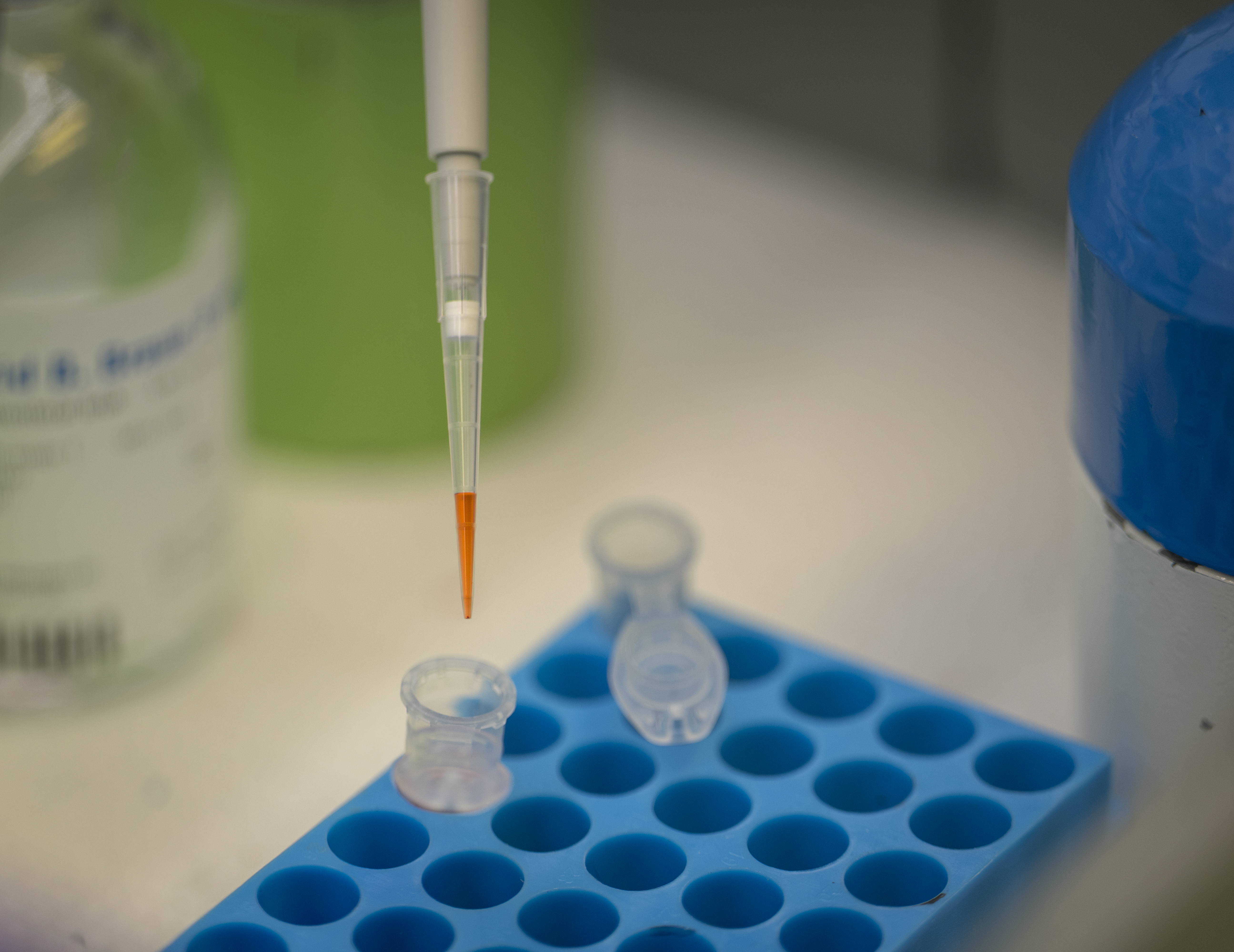
Efforts related to radiotracer development and initial tests to evaluate radioligand binding specificity, affinity, internalization and stability will be conducted in experimental models comprising 2D and 3D cell cultures, organoids, tumor tissue explants as well as other ex vivo models.
In vivo preclinical (mouse and pigs)
A central aim of THERACAN is to integrate imaging and therapy in a way that allows treatment to be monitored and adjusted based on biological response. PET and SPECT imaging are used to quantify radiotracer distribution, enabling assessment of pharmacokinetics, tumor dosimetry, and treatment effect.
The project uses preclinical mouse models of glioblastoma, prostate cancer, breast cancer and other cancer forms, which provide a controlled environment for evaluating therapeutic delivery, imaging accuracy, and treatment response before translation toward clinical applications.