Advanced Microscopy Core Facility
Gallery
Below are some examples of images acquired on microscope systems at AMCF.


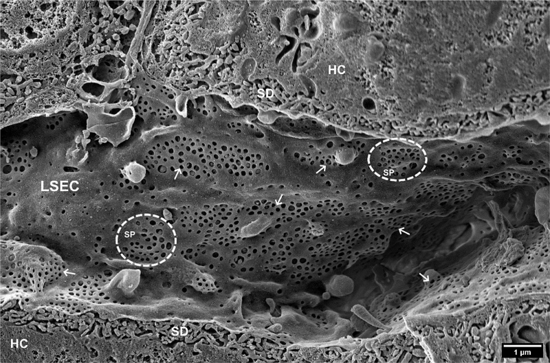
Scanning electron microscopy image of hepatic sinusoids of a C57BL6 mouse, approximately 4 months old. Liver Sinusoidal Endothelial Cells (LSECs) are covered in multiple fenestrations (arrows) arranged into sieve plates (SP, dotted line circles) distributed over the whole sinusoid. SD, space of Disse; HC, hepatocytes. (Courtesy of Karen K. Sørensen, Vascular Biology Research Group). ZEISS Sigma

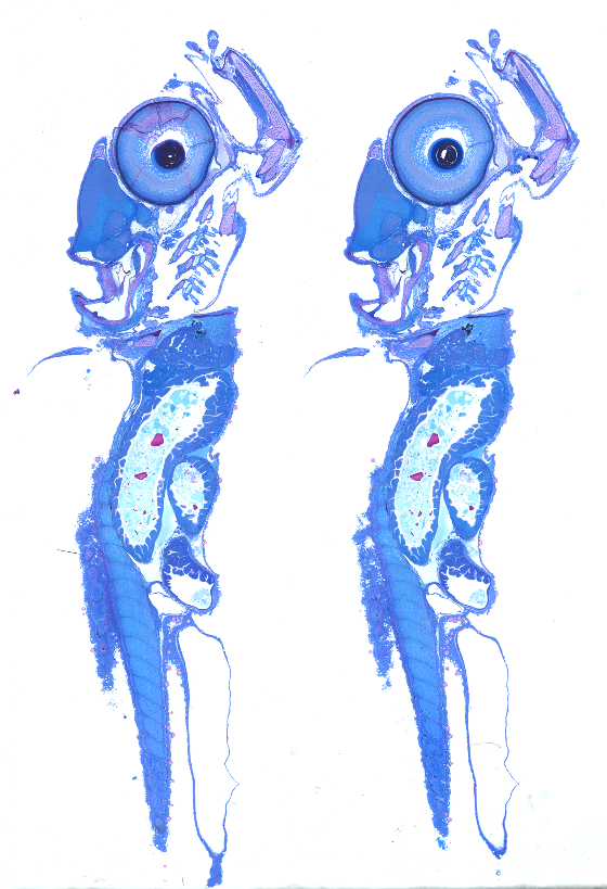
Brightfield microscopy of cod larvae thin section stained with methylene blue-azure II-basic fuchsin. The image is stitched from 72 individual tiles at 112x magnification. ZEISS AxioZoom V.16
Live cell imaging of HEK293 cells using phase contrast. Nikon BioStation IM-Q
Live cell imaging of GFP-tagged LC3B in retinal pigmented epithelial cells using Airyscan FAST. ZEISS LSM880

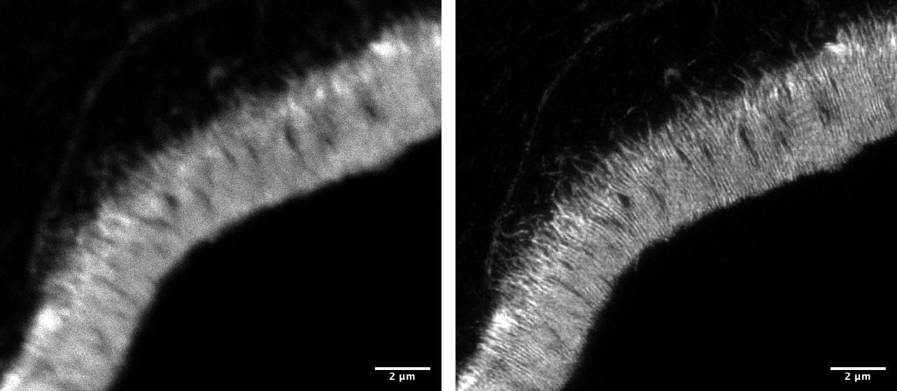
Confocal microscopy (left) versus STED super-resolution (right). ATTO647-phalloidin-labelled cryosection (400 nm thickness) showing microvilli in fish intestine. Abberior STEDYCON